
Journal of Gastroenterology Research and Practice
Case Report - Open Access, Volume 4
Duodenal lipoma: An incidental finding or something more?
Abrol Aradhya, MD1*; Khaira Gurkaramjit, MD2; Kumar Kishore, MD3; Gupta Sorab, MD4; Goyal Abhinav, MD3,6
1Medical Student, Dr Rajendra Prasad Govt Medical College, India.
2Internal Medicine, Geisinger Community Medical Center, USA.
3Division of Gastroenterology, Medicine Institute, Geisinger Clinic, USA.
4Department of Haematology and Oncology, Geisinger Clinic, USA.
6Assistant Professor of Medicine, Geisinger Commonwealth School of Medicine, USA.
*Corresponding Author : Abrol Aradhya, MD
Medical Student, Dr Rajendra Prasad Govt Medical
College, India.
Email: aradhyaabrol98@gmail.com
Received : Dec 29, 2023
Accepted : Feb 09, 2024
Published : Feb 16, 2024
Archived : www.jjgastro.com
Copyright : © Aradhya A (2024).
Abstract
This is a case report of successful endoscopic resection of duodenal lipoma. A 68-year-old male with intermittent vomiting for 1 month. Upper endoscopy revealed a 3 cm pedunculated tumor in horizontal part of duodenum. Endoscopic resection was successful. Final pathological diagnosis was a lipoma. The patient reported complete resolution of symptoms on follow up.
Keywords: Endoscopic resection; Lipoma of duodenum; Duodenal obstruction.
Citation: Aradhya A, Gurkaramjit K, Kishore K, Sorab G, Abhinav G. Duodenal lipoma: An incidental finding or something more?. J Gastroenterol Res Pract. 2024; 4(2): 1185.
Introduction
Lipomas are benign, slow-growing mesenchymal tumors generally found in subcutaneous tissues of proximal extremities and trunk [1]. Gastrointestinal lipomas are uncommon and account for 4 % of all benign tumors. Duodenal lipomas are even rarer. Within the gastrointestinal tract, the colon (64%) is the most common site, followed by the ileum or jejunum (26%), and then the duodenum (4%) [2,3]. Of these, 20% are located in the Duodenal bulb (D1), 50 % are located in descending part (D2), 16 % in the horizontal part (D3), and 9% in ascending part (D4), with 5% unspecified [4]. Peak incidence is around 5th and 7th decade of life. On histology, 90% of GI lipomas are in the submucosa, and 10 % arise from the subserosa [5]. Their shape can be sessile or pedunculated. Most of these tumors are incidentally found and asymptomatic. Here, we report a case of symptomatic lipoma in the horizontal/transverse part of the duodenum that was successfully treated with endoscopic resection.
Case report
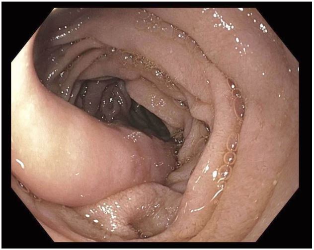
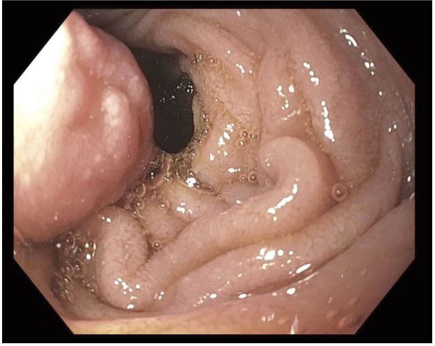
A 68-year-old male with coronary artery disease on aspirin, hypertension, diabetes mellitus on an insulin pump, aortic stenosis status post open aortic valve replacement with porcine valve, Parkinson’s disease on carbidopa-levodopa presented to the office with a 1-month history of intermittent, sudden vomiting without nausea. Episodes of vomiting could occur from 3-4 times per week to several episodes in a single day. He reported vomiting happened half an hour after eating and consisted of the last meal. Vomiting was reported to be projectile. The patient denied any dysphagia, odynophagia, heartburn, regurgitation, or weight loss. No overt bleeding was reported either. The last labs done 2 months before this visit (CBC, CMP) were only remarkable for 122 mg/dL glucose, but no electrolyte abnormality was noted. CT of the abdomen and pelvis with intravenous but no oral contrast done 2 months before this visit had revealed an incidental finding of a duodenal lesion measuring 1.3 cm, which was thought to be a lipoma. He subsequently underwent an upper endoscopy. A 3 cm pedunculated polypoidal nodule was found in 3rd part of the duodenum. Small erosion was noted on the head of this nodule. This nodule was noted to be filling up the entire lumen upon initial examination and half the lumen with maximal insufflation. This nodule was resected using a hot snare, and the mucosal defect was closed using an over-the-scope clip. The patient resumed diet and reported resolution of symptoms in a 2-week follow-up after the procedure. Pathology confirmed the nodule to be a submucosal lipoma.
Discussion
Duodenal Lipomas (DLs) are extremely rare, with incidences limited to case reports. If a duodenal lipoma is symptomatic, the most common findings are gastrointestinal bleeding, ulceration, bowel obstruction, or intussusceptions. Based on the literature review, 80% of symptomatic DLs are larger than 2 cm in diameter. If symptomatic, the most common clinical presentation is small bowel obstruction [6,7] bleeding [8,10] or intussusceptions [11]. The differentiation between a duodenal lipoma and other gastrointestinal tumors, like GIST or liposarcomas, can sometimes be made by CT or MRI. However, an endoscopic ultrasound and fine needle aspiration are sometimes required for diagnosis. On CT, duodenal lipomas can appear as a smooth margined mass with a low Hounsfield unit (range -70 and -120) corresponding with the density of fat [12,13]. As for MRI, the signals are low on T1- and T2-weighted fat-suppressed images, which is specific for lipoma. Lipoma shows no contrast enhancement. Though CT and MRI are helpful in diagnosis, these are unable to locate the origin of the lesion precisely. Direct visualization at endoscopy provides clues about the nature of the tumor. Endoscopy can sometimes be helpful in diagnosis by the appearance of shiny yellow fat while obtaining bite-on-bite biopsies when the mucosa is uncovered, which is sometimes referred to as “the naked fat sign” [14]. Still, it is generally insufficient to make a definitive diagnosis if the lipoma is subserosal. EUS effectively provides information about the layer of origin, echogenicity, and the depth. Asymptomatic duodenal lipomas often do not require any treatment, but symptomatic duodenal lipomas need treatment. Duodenal lipoma can be pedunculated or sessile. The pedunculated lipoma can be safely removed by electrosurgical endoscopic snare polypectomy [6,11,15]. It can also be excised surgically. Endoscopic excision may be challenging and increase the risk of bleeding and perforation for large and sessile lesions. These types of lipomas are challenging to manage using endoscopic techniques; therefore, surgical excision would be the preferred approach. If the nature of the lesion cannot be ascertained or clinical presentation such as intussusception, surgery is required. To our knowledge, this is the first report of a large duodenal lipoma causing symptoms via intermittent duodenal obstruction that was managed endoscopically.
Conflicts of interest: None of the authors declared any conflict of interest, financial or otherwise. No financial support was received in creation of this manuscript.
Acknowledgments: None.
References
- Ouwerkerk HM, Raber, Freling G, Klaase JM. Duodenal Lipoma as a Rare Cause of Upper Gastrointestinal Bleeding. Gastroenterol Res. 2010; 3(6): 290-2.
- O’Riordan BG, Vilor M, Herrera L. Small bowel tumors: an overview. Dig Dis. 1996; 14(4): 245-57.
- Benign small bowel tumor. Accessed September. 2023. Available from: https://www.ncbi.nlm.nih.gov/pmc/articles/PMC1343763/
- Pei MW, Hu MR, Chen WB, Qin C. Diagnosis and Treatment of Duodenal Lipoma: A Systematic Review and a Case Report. J Clin Diagn Res. 2017; 11(7): 01-05.
- Fernandez MJ, Davis RP, Nora PF. Gastrointestinal lipomas. Arch Surg. 1983; 118(9): 1081-3.
- Blanchet MC, Arnal E, Paparel P, Grima F, Voiglio EJ, Caillot JL. Obstructive duodenal lipoma successfully treated by endoscopic polypectomy. Gastrointest Endosc. 2003; 58(6): 938-9.
- Chen HT, Xu GQ, Wang LJ, Chen YP, Li YM. Sonographic features of duodenal lipomas in eight clinicopathologically diagnosed patients. World J Gastroenterol. 2011; 17(23): 2855-9.
- Kadaba R, Bowers KA, Wijesuriya N, Preston SL, Bray GB, Kocher HM. An Unusual Cause of Gastrointestinal Bleeding: Duodenal Lipoma. Case Rep Gastroenterol. 2011; 5(1): 183-8.
- Tung CF, Chow WK, Peng YC, Chen GH, Yang DY, Kwan PC. Bleeding duodenal lipoma successfully treated with endoscopic polypectomy. Gastrointest Endosc. 2001; 54(1): 116-7.
- Zirpe D, Wani M, Tiwari P, Ramaswamy PK, Kumar RP. Duodenal Lipomatosis as a Curious Cause of Upper Gastrointestinal Bleed: A Report with Review of Literature. J Clin Diagn Res. 2016; 10(5): 01-04.
- Knight CD, Black BM. Duodenojejunal intussusception due to lipoma: report of a case. Proc Staff Meet Mayo Clin. 1951; 26(17): 320-3.
- Kakitsubata Y, Kakitsubata S, Nagatomo H, Mitsuo H, Yamada H, Watanabe K. CT manifestations of lipomas of the small intestine and colon. Clin Imaging. 1993; 17(3): 179-82.
- Taylor AJ, Stewart ET, Dodds WJ. Gastrointestinal lipomas: a radiologic and pathologic review. AJR Am J Roentgenol. 1990; 155(6): 1205-10.
- Abu Daff SN, Abu Daff NS. Laparoscopic enucleation of a duodenal lipoma, with review of the literature. Saudi Med J. 2008; 29(3): 455-7.
- Fawcett NW, Bolton VL, Geever EF. Multiple Lipomas of the Stomach and Duodenum. Ann Surg. 1949; 129(4): 524-7.