
Japanese Journal of Gastroenterology Research
Research Article - Open Access, Volume 2
Colonic diverticulosis at colonoscopy in Yaounde (Cameroon)
Ndjitoyap Ndam Antonin Wilson1,2*; Bekolo Nga Winnie3; Awana Armel4; Kenne Yimagou Edgar2; Nsenga Njapa Guy Roger5; Kowo Mathurin Pierre1; Talla Paul1; Ankouane Andoulo Firmin2; Ndjitoyap Ndam Elie Claude2
1Hepatogastroenterology unit, Yaoundé General Hospital, Yaoundé, Cameroon.
2Internal Medicine and Specialities Department, Faculty of Medicine and Biomedical Sciences of the University of Yaoundé I, Yaoundé, Cameroon.
3Clinic Sciences Department, Faculty of Medicine and Pharmaceutic Sciences of the University of Douala, Douala, Cameroon.
4Biomedical Sciences Department, Faculty of Sciences of the University of Ngaoundéré, Ngaoundéré, Cameroon.
5Internal Medicine and Specialities Department, Faculty of Medicine and Pharmaceutic Sciences of the University of Dschang, Dschang, Cameroon.
*Corresponding Author : Ndjitoyap NA Wilson
Hepatogastroenterology unit, Yaoundé General
Hospital, Yaoundé, Cameroon.
Email: tonindam3@yahoo.fr
Received : Apr 29, 2022
Accepted : May 30, 2022
Published : Jun 06, 2022
Archived : www.jjgastro.com
Copyright : © Wilson NNA (2022).
Abstract
Studies about diverticular disease (DD) at colonoscopy are rare in sub-saharan Africa. The aim was to describe the prevalence of the DD at colonoscopy in Yaoundé (Cameroon) regarding the other African studies. We have conducted a retrospective cross sectional study. Recording colonoscopy results from the 1st January 2018 to the 31st December 2021 (4 years). A total of 268 colonoscopies have been performed during the period on 46 patients with diverticulosis (17.2%). The prevalence seems to be slightly higher than in other African countries. The sex ratio was 2.5 (33 men/13 women). The mean age was 63.7 ± 9.6 years (minimum 39 and maximum 80 years-old). Unlikely to what have been described in other African studies, we have observed the same number of right and left colonic localisation with 65.2%. The transverse colon was affected in 52.2%, and a pan diverticulosis in 23.9%. Other significant lesions observed during the colonoscopy were: dolichocolon in 41.3%, polyp in 37%, hemorroid in 34.8%, erosions or ulcerations of the mucosa in 17.4%, coprolithes in 6.5%, and a malignant tumor in 4.4% of cases. The prevalence of the DD at colonoscopies in Yaoundé (Cameroon) is 17.2%. This prevalence seems to be slightly more evaluate than in other African studies. The male sex is more represented. The affection is generally asymptomatic at the diagnosis. All parts of the colon could be affected.
Keywords: Prevalence; Diverticulosis; Colonoscopy; Cameroon.
Citation: Wilson NNA, Winnie BN, Armel A, Edgar KY, Roger NNG, et al. Colonic diverticulosis at colonoscopy in Yaounde (Cameroon). Japanese J Gastroenterol Res. 2022; 2(8): 1086.
Introduction
A diverticulae of the colon is a protusion of its mucosa and submucosa through the muscle layer, which tend to occur at anatomically weak points where blood vessels penetrate the muscular layer [1]. Risks factors evocated are sex, age, constipation, lifestyle (diet, and physical activity), and drugs. The pathophysiology is not completely validated, but various theories are existing: faecal stasis, chronic inflammation, alterations in the intestinal microbiota, neuromuscular alterations, and genetic [2-6]. Diverticulosis is usually detected incidentally on patients undergoing endoscopy or radiological examinations [6]. At the diagnosis, 80% are asymptomatic [7]. But some affected people will develop symptomatic diverticulosis with complicationssuch as acute diverticulitis or diverticular haemorrhage [6]. Publications have showed that diverticulosis are more frequent in the Western [7,8]. But studies available in Africa are rare or old: Archampong et al in Ghana 1978, Segal and walker in South Africa 1982, Madiba and Mokoena in South Africa 1994, KiguliMalwadde and Kasozi in Uganda 2002 [9-12]. Few one recently available have shown the increasing of the prevalence of this affection [1,7]. To know the profile of patients, clinical manifestations and endoscopic features of diverticules of the colon could help to prevent the onset of complications. This is an original study of the diverticular disease (DD) in a Subsaharan Africa country, presenting clinical features of the affection.
Objective
The main objective of our study was to describe the endoscopic profile of diverticules on the colonoscopy in Yaoundé (Cameroon) regarding the results of other African countries.
Methodology
We have conducted a retrospective study from the 1st January 2018 to the 31st December 2021 (4 years) at the Yaoundé General Hospital (Cameroon). The gastroenterologic unit of this hospital has 4 medical doctors specialized in digestive diseases. Each one was performed once a week upper and lower digestive endoscopies. They were using a videoendoscopy device Fujinon® or Storz® model. We have analysed all lower endoscopic reports of patients aged more than 18 years old. We have retained all colonoscopies for which at least one diverticulae has been observed. Repeated exams were excluded. For these coloscopies, we have described the age and the sex of the patient, the indication of the exam, the complete or incomplete character of the colonoscopy, the quality of the colonic preparation respecting the Boston classification (good, middle and poor), the number and the site of diverticules on the colon, the presence of a complication (bleeding) of the diverticule. And we also looked for other lesion associated to diverticules. The study has been approved by the administration of the Yaoundé General Hospital and the ethical committee of the Faculty of Medicine and Biomedical Sciences of the University of Yaounde 1. Data were analysed with SPSS version 20.0.
Results
A total of 268 colonoscopies have been perfomed during the period. At least one diverticulae has been observed in 46 colonoscopies (17.2%). For these patients, men were 33 and women 13, the sex ratio was 2.5 (Table 1). And the mean age was 63.7 ± 9.6 years (minimum 39 and maximum 80 years old). The age group most represented was people aged from 60 to 79 years (Table 1). Indications of the colonoscopy were: lower digestive bleeding in 21/46 patients (45.7%), abdominal pain in 15/46 patients (32.6%), constipation in 7/46 patients (15.2%), colorectal cancer screening in 7/46 patients (15.2%), diarrhea in 4/46 patients (8.7%) and loss of weight in 3/46 patients (6.5%) (Table 2). The colonoscopy was complete for 45 patients (97.8%) and incomplete for one patient (2.2%). The quality of the preparation of the colonoscopy was describe as good among 33 patients (71.7%), middle among 10 patients (21.7%), and poor among 3 patients (6.5%). Diverticular orifices observed were alone in 8/46 patients (17.4%), they were 2 to 5 in 15/46 patients (32.6%) and at least 6 diverticular orifices 23/46 patients (50%). They were presents at the right part of the colon in 30/46 coloscopies (65.2%), transverse part of the colon in 24/46 colonoscopies (52.2%) and left part of the colon in 30/46 coloscopies (65.2%) (Table 2). There were a pan diverticulosis in 11/46 patients (23.9%) (Figure 1). Concerning complications, diverticules were bleeding in 3/46 patients (6.5%) (Figure 2). These were two men of 43 and 80 years-old with a pan diverticulosis, and a woman of 57 yers-old with only one diverticular orifice of the left part of the colon. These diverticules were associated with a dolichocolon in 19/46 patients (41.3%), a polyp in 17/46 patients (37%), hemorroids in 16/46 patients (34.8%), erosions or ulcerations of the mucosa in 8/46 patients (17.4%), coprolithes in 3/46 patients (6.5%), and a malignant tumor in two patients (4.4%) (Table 2).
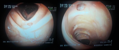
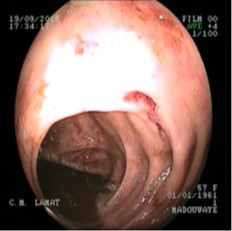
Table 1: Demographic characteristics of patients.
|
Number (n= 46) |
Percentage |
Sex |
33 |
71.7 % |
Age |
1 |
2.2 % |
Table 2: Endoscopic patterns of diverticulosis.
|
Number (n= 46) |
Percentage |
Indication of the colonoscopy |
21 |
45.7 % |
Site of the colon |
30 |
65.2 % |
Number of diverticules |
8 |
17.4 % |
Associated lesions |
16 |
34.8 % |
Discussion
Diverticulosis was observed at 17.2% of colonoscopies. This prevalence is higher than those observed in Nigeria 10.6% in 2016, in South Africa 13.5% in 2017, and in Sudan 7.5% in 2020 [1,7,8]. But the prevalence remains low compared to the West [6]. Our study has revealed a sex ratio of 2.5. Western studies as well as Nigeria and Sudan studies have also described this important predominance of the male sex [1,8]. But the South African research publication has described 27 females against 20 males [7]. They did not explain this female predominance.
The range of 60 to 79 years-old was very represented. As in Western and other african studies, the prevalence of the DD increases with age. Before 40 years, the prevalence is low. An USA study has described a prevalence of 32.6% among individuals aged between 50 – 59 years and 71.4% among those of 80 years-old [6]. But the number of colonoscopy decreases among oldest people because of their limited general condition. The first indication of colonoscopies was a lower digestive bleeding. It is one of the main complications of DD. But in only 3 cases, the diverticulosis was responsible of the bleeding. Regardless the study, the DD in colonoscopy are asymptomatic [13]. In our study, we have observed an equal repartition of diverticules between the right and the left sites (65.2%) and a pan diverticulosis in 23.9%. Our results are different from those observed in South Africa, Sudan and Nigeria which have described a left colonic predominance of the DD [1,7,8]. This difference show the importance to realise a complete exploration of the colon in DD.
We have observed some polyps in 17 patients and a malignant tumour in two patients. Concerning polyps which are benign lesions, they are associated with the diagnosis of asymptomatic diverticulosis [13]. It is not the polyp which has created the diverticulosis, but it has indicated the colonoscopy which led to the diagnosis of DD. The DD does not increase risk of colorectal cancer [14]. But in case of diverticulosis, it is important to exclude another aetiology of the bleeding which can be a malignant tumour [6].
Limitations
Our study have some limitations. Firstly, it is a monocentric study. A multicentric study is more representative of the population. Secondly, it is a retrospective study with risks of missing data.
Conclusion
With a prevalence of 17.2%, the DD seems to be more frequent on colonoscopy realised in Yaoundé (Cameroon) than in other African countries. The male sex is more represented. The affection is generally asymptomatic at the diagnosis. All parts of the colon could be affected.
What is already know on this topic
- Few studies about diverticulosis at colonoscopy exist in sub-Saharan Africa. - The prevalence seem to be higher in western than in subSaharan area. - All parts of the colon could be involved with a prevalence of diverticulosis at the left part.What this study adds
- We have observed a significant prevalence for a sub-Saharan country. - We have also observed a higher prevalence in old people and in men. - We have an equal repartition of diverticular disease between left and right parts of the colon in contrary with western studies.Declarations
Competing interests: The authors declare no competing interests.
Authors’ contributions:
NDJITOYAP NDAM Antonin Wilson, wrote the paper
BEKOLO NGA Winnie, performed the analysis
AWANA Armel, performed the analysis
KENNE YIMAGOU Edgar, collected data
NSENGA NJAPA Guy Roger, performed the analysis
KOWO Mathurin Pierre, other contribution
TALLA Paul, collected data
ANKOUANE ANDOULO Firmin, conceived and designed the analysis
NDJITOYAP NDAM Elie Claude, conceived and designed the analysis.
References
- Oluyemi A, Odeghe E. Diverticular disease at colonoscopy in Lagos State, Nigeria. Niger Med J. 2016; 57: 110.
- Elisei W, Tursi A. The Pathophysiology of Colonic Diverticulosis: Inflammation versus Constipation? Inflamm Intest Dis. 2018; 3: 55-60.
- Maguire LH. Genetic Risk Factors for Diverticular DiseaseEmerging Evidence. J Gastrointest Surg. oct 2020; 24: 2314-7.
- Peery AF, Sandler RS, Ahnen DJ, Galanko JA, Holm AN, Shaukat A, et al. Constipation and a Low-Fiber Diet Are Not Associated With Diverticulosis. Clin Gastroenterol Hepatol. déc 2013; 11: 1622-7.
- Tursi A, Elisei W. Role of Inflammation in the Pathogenesis of Diverticular Disease. Mediators Inflamm. 2019: 1-7.
- Tursi A, Scarpignato C, Strate LL, Lanas A, Kruis W, Lahat A, et al. Colonic diverticular disease. Nat Rev Dis Primer. déc 2020; 6: 20.
- Vally M, Koto MZ, Govender M. An investigation of diverticular disease among black patients undergoing colonoscopy at Dr George Mukhari Academic Hospital, Pretoria, South Africa. South Afr Med J Suid-Afr Tydskr Vir Geneeskd. 2017; 107: 137-9.
- Alnzaer AA, Mohamedahmed AYY, Adam YA, Eltyiep E, Suliman SH. Presentation and anatomical distribution of diverticular disease in four hospitals in Sudan. Pan Afr Med J [Internet]. 2020.
- Diverticular disease in an indigenous African community. 2022.
- Diverticular disease in urban Africans in South Africa - PubMed 2002.
- Haemorrhage--the main presenting feature of diverticular disease of the colon in blacks - PubMed, 2022.
- Diverticular disease of the colon in Kampala, Uganda, 2022.
- Wang F-W, Chuang H-Y, Tu M-S, King T-M, Wang J-H, Hsu C-W, et al. Prevalence and risk factors of asymptomatic colorectal diverticulosis in Taiwan. BMC Gastroenterol. déc. 2015; 15: 40.
- Hong W, Dong L, Zippi M, Stock S, Geng W-J, Xu C, et al. Colonic diverticulosis is not a risk factor for colonic adenoma. Ther Clin Risk Manag. mars 2018; 14: 531-7.