Japanese Journal of Gastroenterology Research
Clinical Image - Open Access, Volume 2
Rectum penetration by an intrauterine device
Jian-Xin Zhang1; Yi Ding2; Wei Liu3,4*
1Department of Gastroenterology, Jingmen Second People’s Hospital, Jingmen, China.
2Department of Gastrointestinal Surgery, Jingmen Second People’s Hospital, Jingmen, China.
3Institute of Digestive Disease, China Three Gorges University, Yichang, China.
4Department of Gastroenterology, Yichang Central People’s Hospital, Yichang, China.
*Corresponding Author : Wei Liu
Institute of Digestive Disease, China Three Gorges
University, 8 Daxue Road, Yichang 443000, China.
Email: liuwei@ctgu.edu.cn
Received : Apr 01, 2022
Accepted : May 04, 2022
Published : May 10, 2022
Archived : www.jjgastro.com
Copyright : © Liu W (2022).
Citation: Jian-Xin Z, Ding Y, Liu W. Rectum penetration by an intrauterine device. Japanese J Gastroenterol Res. 2022; 2(7): 1081.
Description
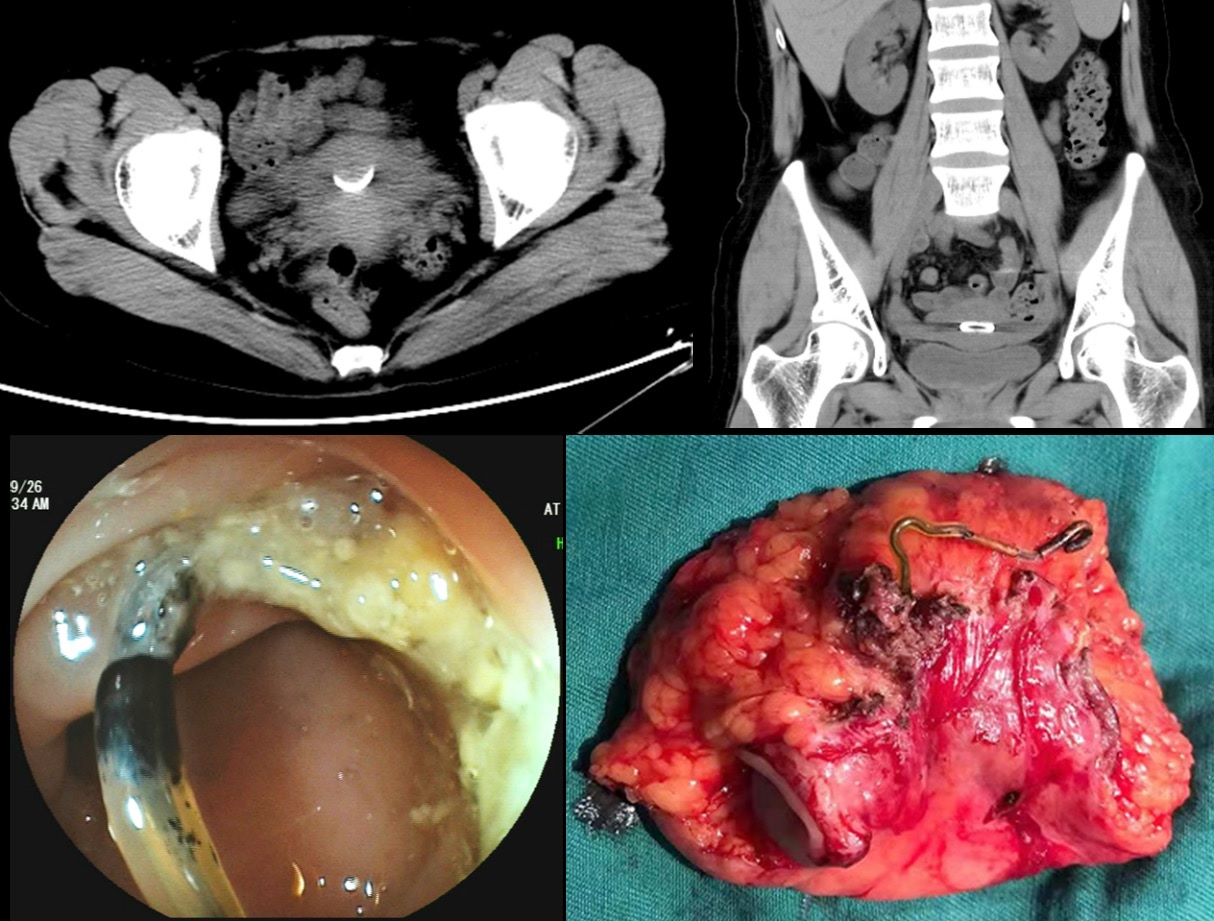
A 38-year-old woman was transferred to our hospital with a 10-day history of lower abdominal pain and vomiting. She had no significant surgical and medical history. Physical examination showed no specific abnormality. Computed tomography of the abdomen and pelvic cavity incidentally showed a thin, irregular foreign object embedded in the right lateral wall of the rectum (Figure 1A and 1B). Colonoscopy confirmed perforation of the rectum by a thin metallic object (Figure 1C). On detailed questioning, she recalled having an intrauterine device inserted 9 years ago. The patient received the diagnosis of rectal perforation by an intrauterine device. The device was successfully removed laparoscopically by performing a partial rectectomy (Figure 1D). The postsurgical course was uneventful and she became asymptomatic after the procedure. As a safe and effective birth control method, insertion of intrauterine contraceptive device is very popular in China. However, the migrated device may present as bleeding, abdominal pain, and even colonic perforation although most of the perforations are asymptomatic [1]. Migrating intrauterine contraceptive devices are usually involved in the sigmoid colon, which is the most commonly penetrated part of the colorectum [2]. As a rare complication of intrauterine device insertions, Uterine perforation may subsequently result in rectal perforation in our case. Minimally invasive techniques such as colonoscopy and laparoscopy are usually performed to remove the device eroding the colon wall [3].
Conflicts of interest: The authors have no conflicts of interest to declare.
Ethical statement: The authors are accountable for all aspects of the work in ensuring that questions related to the accuracy or integrity of any part of the work are appropriately investigated and resolved. Written informed consent was obtained from the patient for publication of this “GI Image”.
Author’s contributions:
Collection of data: Jian-Xin Zhang.
Manuscript preparation and writing: Yi Ding.
Final approval of the manuscript: Wei Liu.
References
- Okamoto T, Fukuda K. Bowel Perforation by Intrauterine Device. Clin Gastroenterol Hepatol. 2021; 19: e79.
- Takahashi H, Puttler KM, Hong C, et al. Sigmoid colon penetration by an intrauterine device: A case report and literature review. Mil Med. 2014; 179: e127-129.
- Peng L, Wei S, Li X, et al. Endoscopic removal of a migrated intrauterine contraceptive device in the rectum assisted by Overstitch defect closure. Endoscopy. 2019; 51: E276-e277.

