
Japanese Journal of Gastroenterology Research
Case Report - Open Access, Volume 2
Lymphoepithelial-like cholangiocarcinoma: A rare primary liver cancer case report
Khue K Dang1; Hoi H Nguyen1; Khiem T Nguyen1*; Tuan H Luong2; Duy V Le1
1Gastrointestinal and Hepato-Biliary-Pancreatic Surgery Department, Bach Mai Hospital, Hanoi, Vietnam.
2General Surgery Department, Hanoi Medical University, Hanoi, Vietnam.
*Corresponding Author : Khiem T Nguyen
Gastrointestinal and Hepato-Biliary-Pancreatic
Surgery Department, Bach Mai Hospital, Hanoi,
Vietnam.
Email: Nguyenthanhkhiemvduc@gmail.com
Received : Feb 28, 2021
Accepted : Apr 07, 2022
Published : Apr 11, 2022
Archived : www.jjgastro.com
Copyright : © Nguyen KT (2022).
Abstract
Introduction: Lymphoepithelioma-like carcinoma is a rare primart liver cancer with unique pathological features. It has characteristics of tumor’s lymphocyte infiltration. It can be classified into lymphoepithelioma-like hepatocellular carcinoma and lymphoepithelioma-like cholangiocarcinoma. It has favourable prognosis in compare with typical hepatocellular carcinoma or intrahepatic cholangiocarcinoma.
Case report: We hereby report case of 71 year-old male patient with chronic hepatitis B infection. He was incidentally discovered a liver mass in left lateral section during routine check-up. The images of the liver tumor has the charecteristics of hepatocellular carcinoma. He underwent laparoscopic left lateral sectionectomy. The final pathological and immunohistochemical result was lymphoepithelioma-like cholangiocarcinoma.
Conclusions: LELC is a rare primary liver cancer, of which the clinical and imaging can be overlap with typical HCC. The diagnosis of LELCC mainly depended on pathological and immunohistochemical result. This is the first case of LEL-CC reported in Vietnam.
Keywords: Liver cancer; Lymphoepithelial-like carcinoma; Cholangiocarcinoma; Case report.
Abbreviations: AFP: Alpha-Fetoprotein; CT: Computed Tomography; HBV: Hepatitis B; HCC: Hepatocellular Carcinoma; ICC: Intrahepatic Cholangiocarcinoma; LELC: Lymphoepithelial-like Carcinoma; LEL-HCC: Lymphoepithelial-Like Hepatocellular Carcinoma; LEL-CC: Lymphoepithelial-Like Cholangiocarcinoma.
Citation: Dang kk, Nguyen HH, Nguyen KT, Luong TH, Le DV. Lymphoepithelial-like cholangiocarcinoma: A rare primary liver cancer case report. Japanese J Gastroenterol Res. 2021; 2(6): 1073.
Introduction
Hepatocellular carcinoma (HCC) and intrahepatic cholangiocarcinoma (iCC) are two most common primary liver cancer. Lymphoepithelial-like carcinoma (LELC) is a very rare type primary cancer of the liver. It has distinctive histological characteristic with sclerotic tissue and lymphatic infiltration. Generally, it was reported that patient with LELC has favourable prognosis in compare with typical HCC or iCC. To date, only 104 cases of both hepatocellular (LEL-HCC), cholangiocellular (LEL-CC) or mixed types have been reported, of which only 34 cases was represented with cholangiocellular type [1].
Case presentation
We report a case of 71 year-old male patient admitted to our department in 10th January 2022. He had history of cirrrhosis and chronic HBV infection treated with TDF. He was incidentally discovered a liver mass during routine check-up and did not have any symptom during hospitalization. He had normal blood counts test and liver function with slightly elevated in liver enzyme (AST: 55U/l, ALT: 50U/l). His Alpha-fetoprotein level was within normal limit: 2,4ng/ml. His upper and lower gastrointestinal endoscopy scanning were normal, without varices.
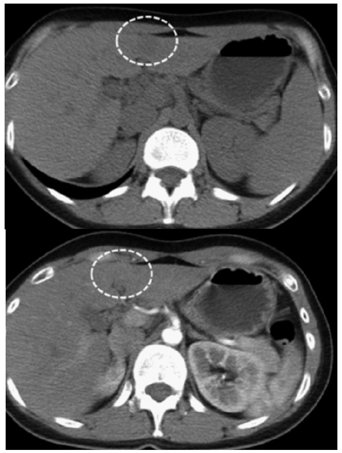
The CT scanner revealed a 2.7 cm hypodense tumor in left lateral segment, with hyperenhancement in arterial phase and wash-out in veinous phase (Figure 1). He was diagnosed with hepatocellular carcinoma and underwent laparoscopic left lateral segmentectomy. The tumor was located in the central of segment 3. The spicimen was shown in (Figure 2). The postoperative course was uneventful, and the patient was discharged on postoperative day 5.
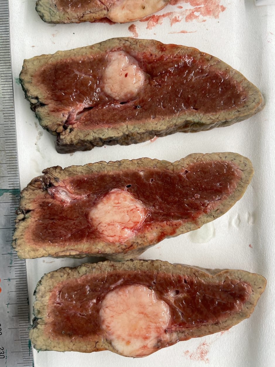
The gross pathology showed a 3.5 cm tumor with uncleared margin. The microscopic pathological of the tumor was consisted of sclerotic tissue with lymphocytes infiltration, the background liver was cirrhosis grade 4 according to METAVIR. The immunohistochemistry of the tumor were positive with CK7, CK19, EMA staining, and negative to CD56, Heppar1, Glypican 3, CK20, TTF1, SATB. The morphological and immunohistochemical result yielded Lymphoepithelial-like cholangiocarcinoma, a rare primary liver neoplasm.
Discussion
LELC is a rare primary liver cancer with its own features, and it was recognized by WHO as a distinctive liver cancer in 2010[2]. LELC originally used to describe a type of nasopharyngeal cancer, and often associated with Epstein-Barr virus (EBV) infection. The first report of primary liver LELC was back from 1995 [3], until now, 104 cases of LELC, of which only 34 LEL-CC type have been reported [1].
Hepatocellular carcinoma is the most common primary liver cancer in the world, and in our country. Our patient hospitalized with HCC-like characteristics. He had a background of cirrhosis with chronic HBV infection. The radiologic features were also mimic HCC, with wash-out features. No further biopsy was necessary, the patient had the criteria for a diagnosis of HCC according to the American Association for the Study of Liver Diseases Practice Guidelines [4]. The laparoscopic liver resection indication was reasonable at the time of clinical diagnosis. The final diagnosis of LELC was solely depended on pathology.
Previous reports also showed that, most patients with LELC had chronic hepatitis infection [5-8]. Chronic hepatitis is also the risk factor for intrahepatic cholangiocarcinoma. Our case also had chronic HBV, however, we still cannot check his EBV infection status. To our knowledge, this is the first case LELC of the liver ever reported in Vietnam.
In term of prognosis, LEL-CC seems to have better long term prognosis than typical cholangiocarcinoma, although the information with LEL-CC is limited because most of the studies were retrospectives case series [7]. The favourable prognosis could be explained due to immune response with lymphocyte infiltration of the tumor.
Conclusions
LELC is a rare primary liver cancer, of which the clinical and imaging can be overlap with typical HCC. The diagnosis of LELCC mainly depended on pathological and immunohistochemical result. This is the first case of LEL-CC reported in Vietnam.
References
- Zhang K, Tao C, Tao Z, et al. Lymphoepithelioma-like carcinoma in liver not associated with Epstein-Barr virus: a report of 3 cases and literature review. Diagn Pathol. 2020; 15: 115 2020.
- Bosman FT. WHO Classification of Tumours of the Digestive. ed 4. Geneva: WHO Press; 2010.
- Shirabe K, Matsumata T, Maeda T, Sadanaga N, Kuwano H, Sugimachi K. A long-term surviving patient with hepatocellular carcinoma including lymphocytes infiltration a clinicopathological study. Hepatogastroenterology. 1995; 42: 996-1001
- Bruix J, Sherman M. American Association for the Study of Liver Diseases. Management of hepatocellular carcinoma: an update. Hepatology. 2011; 53: 1020-1022.
- Ortiz MR, Garijo G, Adrados M, López-Bonet E, Acero D, Bernadó L. Epstein-Barr Virus-Associated Cholangiocarcinoma with Lymphoepithelioma-Like Component. Int J Surg Pathol. 2000; 8: 347-351.
- Jeng YM, Chen CL, Hsu HC. Lymphoepithelioma-like cholangiocarcinoma: an Epstein-Barr virus-associated tumor. Am J Surg Pathol. 2001; 25: 516-520
- Chan AW, Tong JH, Sung MY, Lai PB, To KF. Epstein-Barr virusassociated lymphoepithelioma-like cholangiocarcinoma: a rare variant of intrahepatic cholangiocarcinoma with favourable outcome. Histopathology. 2014; 65: 674-683.
- Gearty SV, Al Jurdi A, Pittman ME, Gupta R. An EBV+ lymphoepithelioma-like cholangiocarcinoma in a young woman with chronic hepatitis B. BMJ Case Rep. 2019; 12: e229520.