
Japanese Journal of Gastroenterology Research
Research Article - Open Access, Volume 2
Anatomical classification of catterpillar hump of RHA and its surgical importance (Nagpur classification)
Prashant Rahate1*; Zoeb Haidar2; Akshay Bangde3; Amol Belsare3; Vipul Golchha3; Kunal Yadav3
1 Department of Minimal Access Surgery, Seven Star Hospital, Nagpur, Maharashtra, India.
2 FRCS, Consultant Surgeon, Director, Seven Star Hospital, Nagpur, India.
3 MUHS Fellow in Minimal Access Surgery, Seven Star Hospital, Nagpur, India.
*Corresponding Author : Rahate PV
Department of Minimal Access Surgery, MS,
CMD, Seven Star Hospital, Nagpur, Maharashtra,
India.
Email: prashantrahate84@yahoo.com
Received : Dec 14, 2021
Accepted : Jan 24, 2022
Published : Jan 31, 2022
Archived : www.jjgastro.com
Copyright : © Rahate PV (2022).
Abstract
Background: Ligation of cystic artery is important surgical step involving gallbladder and hepatobiliary surgery. Right hepatic artery may come very close to gallbladder & cystic duct and CHD in the form of “Caterpillar hump or Moynihan hump’’. Such hump has variations in position and depending on hump type, cystic artery anatomy is defined. In this situation right hepatic artery is liable to be mistakenly identified as cystic artery and it will be ligated prior to Cholecystectomy leading to right functional lobe of liver goes for necrosis. By defining types, increasing surgeon’s awareness, surgical complications will be reduced.
Materials and methods: 600 videos of laparoscopic surgery of gall bladder and CBD exploration were retrospectively reviewed for presence of caterpillar hump in RHA in Rahate Surgical hospital and Sevenstar Hospital, Nagpur, Maharashtra, India from 2012 to 2021 April. Lot of literature was reviewed. Type of hump and its anatomical relations and difficulty level of laparoscopic surgery because of hump was assessed.
Result: Caterpillar hump was present in 21 cases (3.5%) in present study. We found lot of anatomical variations of hump, and judged the level of difficulty of laparoscopic cholecystectomy depending on type of caterpillar hump. We propose a simple classification of type of caterpillar hump depending on observations.
Conclusion: Knowing the vascular anatomy and likelihood of complications should be known to all surgeons. So that the surgeons are able to identify this arterial variation during their cholecystectomy surgeries. If this caterpillar hump of right hepatic artery is present, the surgeons should locate the origin of cystic artery to avoid any unnecessary confusion between cystic artery and right hepatic artery for preventing unnecessary damage to the right hepatic artery. In an attempt to classify caterpillar hump, we can define, predict position of cystic artery type and variation, thereby helping in preventing vascular complications during laparoscopic cholecystectomy and CBD exploration.
Keywords: caterpillar hump; moynihan hump; right hepatic artery injury; cystic artery; cholecystectomy; laparoscopic cholecystectomy; vascular injury during lap; cholecystectomy; difficult callots anatomy
Citation: Rahate PV, Haidar Z, Bangde A, Belsare A, Golchha V, Yadav K. Anatomical classification of catterpillar hump of RHA and its surgical importance (Nagpur classification). Japanese J Gastroenterol Res. 2022; 2(2): 1056.
Introduction
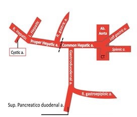
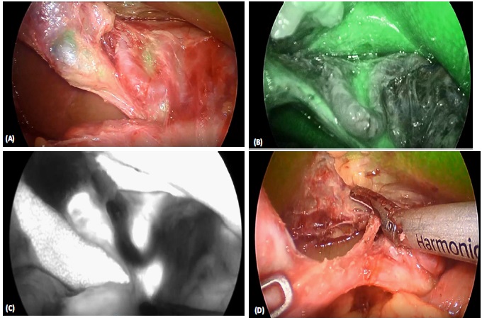
Good optics and infrared imaging added recently in armamentarium of surgeons has increased safety of laparoscopic cholecystectomy and CBD exploration. In spite of advancement and improvement in techniques, certain anatomical variations spring surprises, and may lead to surgical complications -biliary and vascular. Laparoscopic cholecystectomy makes it mandatory to have a thorough knowledge of normal anatomy and variations in this region to reduce the likelihood of uncontrolled intraoperative bleeding, iatrogenic extrahepatic biliary injury and conversion to open cholecystectomy [1]. Incidence of conversion to open surgery because of vascular injury is 0–1.9% and mortality is 0.02% [2]. The right hepatic artery after its origin from hepatic artery proper crosses anterior to the portal vein and then passes behind the common hepatic duct to enter the Calot’s triangle (bounded by cystic duct, common hepatic duct and lower edge of the liver). As it approaches the cystic duct, it gives off the cystic artery and then turns upwards, behind (and between) the right hepatic and the cystic duct to the right lobe of the liver. The cystic artery normally arising from the right hepatic within the triangle, passes in the triangle toward the neck of gall bladder where it typically divides into two branches one of which runs on the attached surface of the gall bladder and the other on its peritoneal surface [3]. Tortuous right hepatic artery, running with upward and downward course producing hump is rarely described anomaly [4]. This tortuosity of the right hepatic artery is called caterpillar hump or Moynihan’s hump [5]. Both inside and outside the Calot’s triangle, the right hepatic often makes a characteristic caterpillar like loop, convexity of which points downward, upward, to the right or to the left. In cholecystectomy, such tortuosity of the right hepatic is extremely vulnerable, for the cystic artery may arise from the distal or the proximal end of the loop, in the latter instance crossing it.
The U shaped right hepatic artery has various positions with reference to the cystic duct.
(A) More nearer the hump to CD shorter will be CA.
(B) The hump can be anterior or posterior to CHD.
(C) Hump can be single loop or double loop.
(D) Cystic artery can be single or double (anterior and posterior).
A bend in the course of the right hepatic artery throwing it into the caterpillar hump invites injury unless it is carefully dissected free [12]. This variant of right hepatic artery invariably leads to abnormalities of cystic artery formation which can result in its injury during surgical procedures.
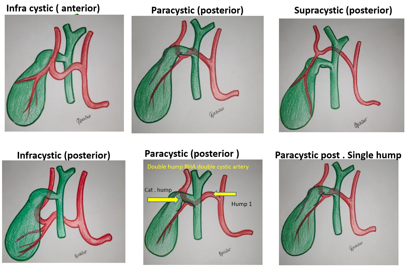
Types of caterpillar hump
Since cystic duct is taken as reference, depending on the anatomical relationship of caterpillar hump to cystic duct classification of hump is done.
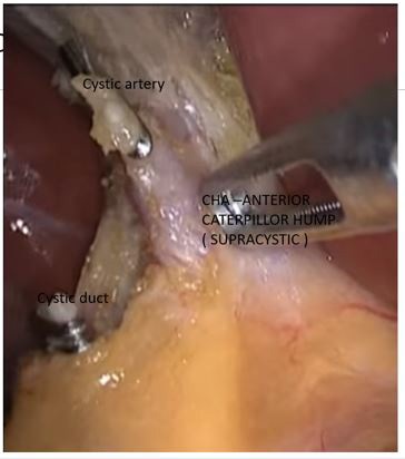
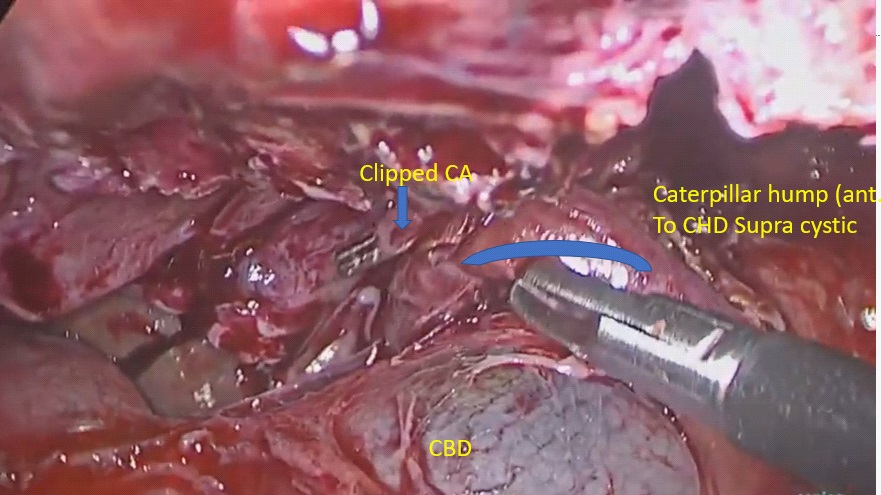
1) Supracystic: The hump is either anterior or posterior to CHD, superior to the cystic duct, nearer to hilum of liver. Here the cystic artery is long, originating from inferior part of loop hump. Due to long cystic artery, and hump remaining away from surgical dissection, mishaps are less during surgery
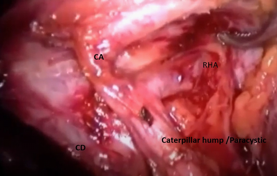
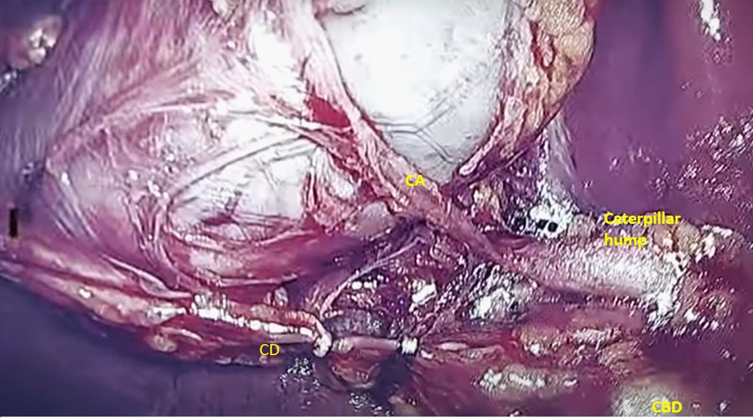
2) Paracystic: The hump is around the confluence of cystic artery and CHD. Here the hump usually gives off two branches, anterior cystic artery and post cystic artery. since the hump is very close to cystic duct, the cystic arteries are very short. the hump (RHA) is mistaken for CA and high chances of vascular catastrophe during surgery.
3) Infracystic: The hump is below the confluence towards duodenum, here again the CA is long, single, originating from ascending part of hump. surgical mishaps are less for obvious reason.
Depending on other anatomical details caterpillar hump can be classified further as follows:
a. Anterior / posterior (loop position in relation to CHD / CBD).
b. Single hump / double hump (depending on number of hump) [8].
c. single cystic artery / double cystic artery.
Surgical significance
Variant of right hepatic artery invariably leads to abnormalities of cystic artery formation which can result in its injury during surgical procedures Since the cystic artery arising from the loop is typically short, it may get easily avulsed from the hepatic artery, if excessive traction is applied to the gall bladder producing brisk bleeding [24].
Sometimes tortuous right hepatic artery does not give a single cystic artery but supplies the gall bladder with several small twigs. The right hepatic artery is injured while securing them. Injury to right hepatic artery can be fatal in presence of impaired liver function [24-26]. It is usually the right hepatic artery that is in danger during this surgery and must be located before ligating the cystic artery [5]. Because numerous variations in origin and branching pattern of right hepatic artery have been reported [27].
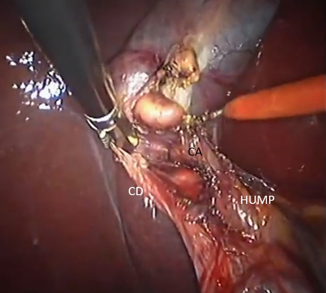
The Hump lies in close proximity to the gall bladder and cystic duct and so it may be mistaken to be cystic artery and inadvertently ligated during surgical procedures like cholecystectomy and liver transplantation [5,7,11,13]. So caterpillar hump should be suspected when an unusually large ‘cystic artery’ is seen through the laparoscope [5,12]. It must be emphasized that an artery resembling the cystic artery in its course and paralleling the cystic duct is not necessarily the cystic artery but may be right hepatic artery and the calibre of vessels to be divided is not a r eliable index of whether it is cystic artery or right hepatic artery. Therefore, it is essential to visualize right hepatic artery above and below the origin of cystic branch [28,29].
Conclusion
With increasing incidence of gall bladder and ductal disease, knowing the vascular anatomy and likelihood of complications should be known to all surgeons. Ductal anatomy had been extensively studied and documented. Vascular anatomy has been little neglected, this is an attempt to make surgeons aware of callots triangle vascular anatomy. So that the surgeons are able to identify this arterial variation during their cholecystectomy surgeries. If this caterpillar hump of right hepatic artery is present, the surgeons should locate the origin of cystic artery to avoid any unnecessary confusion between cystic artery and right hepatic artery for preventing unnecessary damage to the right hepatic artery. In an attempt to classify caterpillar hump, we can define, predict position of cystic artery type and variation, thereby helping in preventing vascular complications during laparoscopic cholecystectomy and CBD exploration. This is single centre observation involving a single surgical team. This topic needs multi centre analysis with surgeons and anatomist.
References
- Zubair M, Habib L, Mirza MR, et al. Anatomical variations of cystic artery: telescopic facts. Med J Malaysia. 2012; 67: 494-496.
- Ding YM, Wang B, Wang WX, et al. new classification of the anatomical variations of cystic artery during laparoscopic cholecystectomy. World J Gastroenterol. 2007; 13: 5629-563.
- Holinshed WH. The Liver and Gall bladder. Anatomy for Surgeons. The Thorax Abdomen and Pelvis. vol. 2. New York: Harper and Brothers. 1956; 346-347.
- Jansirani D, Mugunthan N, Phalgunan V, et al. Caterpillar hump of right hepatic artery: incidence and surgical significance. Natl J Clin Anat. 2012; 1: 121-124.
- Michel’s NA. Blood Supply and Anatomy of the Upper Abdominal Organs with Descriptive Atlas. Philadelphia: Lippincott Company; 1955: 171-175.
- Hamza MU, Jaffar AA, Hassan HA, et al. Vascular and gall bladder variations in laparoscopic cholecystectomy. Med J Babylon. 2008; 5: 119-130.
- Johnston EV, Anson BJ. Variations in the formation and vascular relationships of bile ducts. Surg Gynecol Obstet. 1952; 94: 669- 686.
- Kavitha B. An anatomical study of Moynihan’s hump of right hepatic artery and its surgical importance, J Anat Soc India, 2016.
- Bergamaschi R, Ignjatovic D. More than two structures in Calot’s triangle: a post-mortem study. Surg Endosc. 2000; 14: 354-357.
- Ayyaz M, Fatima T, Ahmed G. Arterial anatomy in Calot’s triangle as viewed through the laparoscope. Ann King Edward Med Coll. 2001; 7: 183-185.
- Al-Sayigh HA. The incidence of cystic artery variation during laparoscopic surgery. Med J Babylon. 2010; 7: 389-403.
- Mishall PL, Rajgopal L. Variant right hepatic artery forming Moynihan’s hump – clinical relevance. Int J Anat Var. 2010; 3: 144-145.
- Bhargava GS, Singh H, Singh HD, et al. Moynihan’s hump of right hepatic artery: a case report and surgical significance. CIBTech J Surg. 2014; 3: 42-44.
- Bagadabettu SN, Sirsangandla SR, Kumar N, et al. Hepatosplenic trunk associated with tortuous course of right hepatic artery forming caterpillar hump. N Am J Med Sci. 2012; 4: 376-378.
- Bulut T, Yamaner S, Bugra D, et al. False aneurysm of the hepatic artery after laparoscopic cholecystectomy. Acta Chir Belg. 2002; 102: 459-463.
- Alves A, Farges O, Nicolet J, Watrin T, Sauvanet A, Belghiti J: Incidence and consequence of an hepatic artery injury in patients with post cholecystectomy bile duct strictures. Ann Surg 2003; 238: 93-96.
- Halasz NA: Cholecystectomy and hepatic artery injury. Arch Surg 1991; 126: 137-138.
- Chapman WC, Halevy A, Blumgart LH, Benjamin IS: Post-cholecystectomy bile duct strictures: management and outcome in 130 patients. Arch Surg 1995; 130: 597-604.
- Deziel DJ, Millikan KW, Economou SG Doolas A, Ko ST, Airan MC: Complications of laparoscopic chomesystectomy: a national survey of 4,292 hospials and an analysis of 77,704 cases. Am J Surg 1993; 165: 9-14.
- Bismuth H: How to treat a postoperative stenosis? in Bismuth H, Lazorthes F (eds): Operative Injury of the Common Bile Duct. Paris, Masson. 1981; 47-107.
- Nicholson T, Travis S, Ettles D, Dyet J, Sedman P, Wedgewood K, Royston C: Hepatic artery angiography and embolization for haemobilia following laparoscopic cholecystectomy. Cardiovasc Intervent Radiol. 1999; 22: 20.
- Connor S, Garden OJ: Bile duct injury in the era of laparoscopic cholecystectomy. Br J Surg 2006; 93: 158-168.
- Frilling A, Li J, Weber F, Frubauf NR, Engel J, Beckebaum S, et al. Major bile duct injuries after laparoscopic cholecystectomy: a tertiary center experience. J Gastrointest Surg 2004; 8: 679-685.
- Al-Sayigh HA. The incidence of cystic artery variation during laparoscopic surgery. Med J Babylon. 2010; 7: 389-403.
- Jansirani D, Mugunthan N, Phalgunan V, et al. Caterpillar hump of right hepatic artery: incidence and surgical significance. Natl J Clin Anat. 2012; 1: 121-124.
- Bhargava GS, Singh H, Singh HD, et al. Moynihan’s hump of right hepatic artery: a case report and surgical significance. CIBTech J Surg. 2014; 3: 42-44.
- IKeith L. Moore, clinically oriented anatomy, 6th edition, 2009; 287-288.
- Hamza MU, Jaffar AA, Hassan HA, et al. Vascular and gall bladder variations in laparoscopic cholecystectomy. Med J Babylon. 2008; 5: 119-130.
- Variant right hepatic artery forming Moynihan’s hump: clinical relevance, L Rajgopal - Int J Anat Var, 2010.