
Japanese Journal of Gastroenterology Research
Case Report - Open Access, Volume 1
Ovarian torsion in strangulated inguinal hernia; A rare complication with a high morbidity: Case report
Ahmed Elrouby
Associate Professor of Pediatric Surgery, Faculty of Medicine, Alexandria University, Egypt.
*Corresponding Author : Ahmed Elrouby
Associate Professor of Pediatric Surgery, Faculty of
Medicine, Alexandria University, Egypt.
Email: elroubypaedo@yahoo.co
Received : Oct 20, 2021
Accepted : Dec 03, 2021
Published : Dec 07, 2021
Archived : www.jjgastro.com
Copyright : © Elrouby A (2021).
Abstract
The incidence of congenital inguinal hernia ranges between 0.8% and 4.4% being more common in males than in females and mostly it is indirect. The ovary is commonly prolapsed in female inguinal hernia, however ovarian torsion is uncommon. I report a five month old female patient with an ovarian torsion in a strangulated inguinal hernia with its investigatory findings and her proper management.
Keywords: strangulated; ovarian torsion; inguinal hernia.
Citation: Elrouby A. Ovarian torsion in strangulated inguinal hernia; A rare complication with a high morbidity: Case report. Japanese J Gastroenterol Res. 2021; 1(9): 1042.
Introduction
Congenital inguinal hernia is the most common surgical problem faced in pediatric surgery. A prompt diagnosis with urgent intervention is essential to abort the risk of complications which associate this surgical problem [1]. Although the ovary is commonly prolapsed in female congenital inguinal hernia; it is uncommon to be complicated with torsion [2]. So we report a five month old female patient presented with a strangulated left inguinal hernia with ovarian torsion in association with the investigatory findings and the management done.
Case report
I present a five month old female patient who presented to our institute with her parents complaining of a left inguinal swelling since two days in association with severe crying resulting from agonizing pain developing from the same swelling. Vomiting developed two times with a yellowish content since one day. General examination revealed an average body weight without any alarming change in the vital signs. Local inguinal examination revealed a solitary swelling about 3 X 3 cm, rounded in shape, with no impulse on crying and with normal skin overlying. Palpation showed an irreducible tense swelling with severe local tenderness. The swelling did not reveal impulse on crying; also negative fluctuation and trans-illumination tests were elicited.
US examination showed a left inguinal hernia along the canal of Nuck admitting the left ovary which is relatively increased in size without showing any internal vascularity on continuous color Doppler sonographic examination. Laboratory investigations did not show any abnormality except for leukocytosis with neutrophilia.
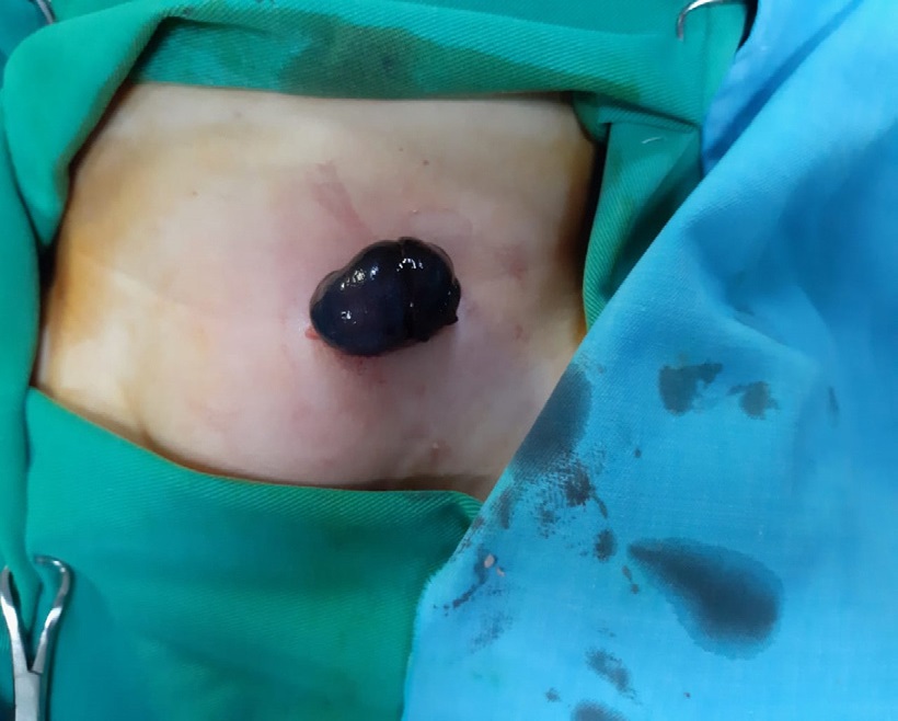
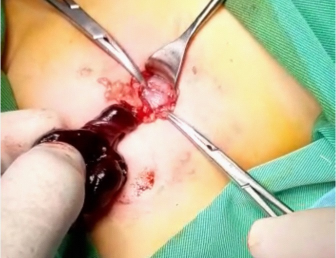
After a written consent was signed by the parents with complete explanation of the possibility of oophorectomy; an immediate left inguinal exploration under general anesthesia with endotracheal intubation was done. Opening of the Camper’s and Scarpa’s fasciae revealed massive tissue edema with friable bleedy tissues. Delivery of the hernia sac with its content was done through the wound followed by cautious opening of the sac with extrusion of sangunious collection followed by the appearance of a gangrenous friable ovary with two rounds of anticlock wise torsion around its vascular pedicle (Figure 1 & 2).
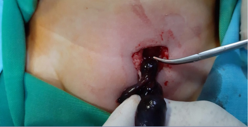
Immediate de-torsion of the ovary was done showing a severely congested friable ovarian tissue with multiple points of damage affecting its wall with severely injured friable medullary tissue. Opening of the external oblique aponeurosis was then done followed by hot fomentations of the ovary for about 15 minutes with 100% O2 supply without showing any improvement in the ovarian condition. Also snipping of the ovarian wall with the needle did not show any bleeding so the decision of oophorectomy was done followed by good hemostasis (Figure 3 & 4).
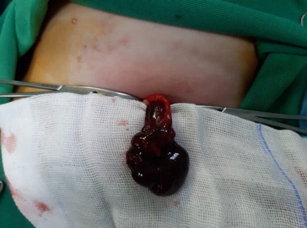
Hernia sac was totally dissected from the surroundings followed by its transfixtion- ligation using Vicryl 3/0. Posterior inguinal wall repair was done using the same suture material followed by closure of the inguinal region in layers. The patient recovered smoothly and passed the post-operative period without any complications. The specimen was sent for pathological examination and revealed gangrenous ovary.
Discussion
The inguinal hernia is one of the most common surgical problems faced in pediatric surgery. It results from persistence of patent processus vaginalis in males and the canal of Nuck in females. The content of the hernia sac may be the intestine, the omentum, a maldescended testis, the ovary and or a sliding hernia with the urinary bladder lying in the sac [3].
The incidence of ovarian incarceration in female inguinal hernia is about 15-30%. On the other hand; ovarian torsion with or without the fallobian tubes can develop in 2-43% of inguinal hernias. Adnexal torsion whether with or without hernia may be in the form of isolated fallopian tube torsion or in association with ovarian torsion. Isolated tubal torsion develops when the tube is prolapsing in hernia sac while the ovary is normally fixed in its place what is called the bell-clapper deformity which is similar to the pathology in testicular torsion [2].
Ovarian torsion develops easily when it is prolapsing in hernia sac with inadequate fixation due to the fact that the ovarian pedicle is narrow at the deep inguinal ring with a relatively larger ovarian size than the pedicle so predisposing the ovary easily for torsion. This fact imposes an urgent surgical intervention in case of ovarian prolapse in hernia sac due to the relatively narrow pedicle to avoid losing a reproductive organ like the ovary or the fallopian tube [2].
Unlike testicular necrosis in case of strangulated hernia in male-which results from vascular compromise caused by the surrounding intestinal loops- ovarian necrosis develops in case of ovarian torsion in strangulated hernia at its narrowed pedicle by lymphatic obstruction followed by venous obstruction and lastly arterial obstruction and consequently if urgent management did not take place immediately; necrosis will be un avoidable. So the presence of an irreducible hernia in female child with tender mass should raise the possibility of incarcerated organ mostly the ovary in an inguinal hernia which requires an urgent intervention [4]. Sonographic examination in association with Doppler US have to be carried in such situations not only to confirm the diagnosis but also to examine the viability of the strangulated organ [5]. For example when the hernia sac contain an un-complicated ovary, the ovary is easily palpable in the inguinal canal and is usually reduced easily. In this condition US will reveal a solid mass which is hypoechoic to the surrounding fat with anechoic follicles and good vascularity [5].
On the other hand; a complicated hernia shows tender irreducible ovary showing an increased ovarian size with a heterogenous echo structure in association with the presence of multiple cysts located at the periphery. Absent blood flow with whirlpool sign ; one of the characteristic signs of vascular compromise in case of ovarian torsion [6].
Conclusion
Female congenital inguinal hernia with a prolapsing ovary is considered a true surgical emergency and prompt immediate surgical intervention to protect against losing a reproductive organ like the ovary or fallopian tube in case of torsion.
Declarations
Financial support: There is no financial support.
Conflict of interest: There is no conflict of interest to declare.
Informed consent: An informed consent was signed by the parents accepting publication of this case report.
Previous presentation: The study was neither presented in any meeting nor submitted for publication in other journals.
References
- Helal AA. Inguinal Hernia in Infancy and Children. In: Hernia. InTech. 2017.
- Volkan Sarper Erikci, Cemal Bilir, Ali Sayan TÖ and GK. Ovarian Torsion Associated with Strangulated Inguinal Hernia: A Case Report and Revıew of Lıterature. EC Paediatr Rep. 2018; 478-482.
- Shehata S, Shehata S, Wella HL, Abouheba M, Elrouby A. Pediatric inguinal hernias, are they all the same? A proposed pediatric hernia classification and tailored treatment. Hernia. 2018; 22(6): 941-946.
- Rees MA, Squires JE, Tadros S, Squires JH. Canal of Nuck hernia: a multimodality imaging review. Pediatr radiol.
- Holley A. Pathologies of the canal of Nuck. Sonography. 2018; 5(1): 29-35.
- Aydin S, Ergün E, Fatihoğlu E, Durhan G, Koşar PN. Ovarian torsion within an incarcereted inguinal hernia; Ultrasound and color Doppler findings. Cumhur Med J. 2017; 39(1): 449-449.