Japanese Journal of Gastroenterology Research
Clinical Image - Open Access, Volume 1
Hepatic portal vein gas in gastric outlet obstruction
Kan Radeesri1; Suphakarn Techapongsatorn2,*
1Department of Radiology, Faculty of Medicine, Vajira hospital, Navamindradhiraj University, Bangkok, Thailand.
2Department of Surgery, Faculty of Medicine, Vajira hospital, Navamindradhiraj University, Bangkok, Thailand.
*Corresponding Author: Suphakarn Techapongsatorn
Department of Surgery, Faculty of Medicine, Vajira
Hospital, Navamindradhiraj University, 681 Samsen road, Dusit, Bangkok, 10330, Thailand.
Tel: +66-244-3282
Email: suphakarn@nmu.ac.th
Received : Aug 05, 2021
Accepted : Sep 09, 2021
Published : Sep 14, 2021
Archived : www.jjgastro.com
Copyright : © Techapongsatorn S (2021).
Clinical image description
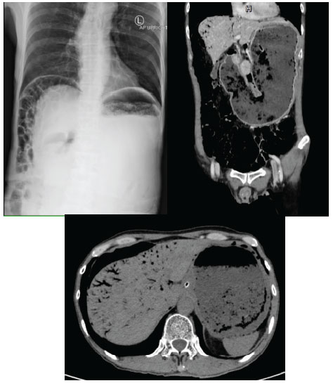
A 55-year-old man presented to an emergency department with a history of abdominal pain and vomiting for one week. He had a history of having peptic ulcer perforation surgery. He appeared weak and frustration from pain, abdominal distension at upper abdomen without peritonitis sign on physical examination. Initial abdominal radiograph revealed pneumoperitoneum under both hemidiaphragms with markedly distension of stomach containing food content. Further computed tomography demonstrated evidence of gastric outlet obstruction without intra or extraluminal mass. There is also massive amount of portal venous gas in both lobes of liver. After patient resuscitation with intravenous fluid and nasogastric intubation for gastric decompression, his condition returned to normal, with no sign of peritonitis nor sepsis. Therefore, the upper gastrointestinal endoscopy showed gastric outlet obstruction from chronic peptic ulcers. The endoscopic balloon dilatation of the obstruction part was successful, and he was discharged home with full recovery in one week.

