
Japanese Journal of Gastroenterology Research
Case Report - Open Access, Volume 1
May-Thurner syndrome successfully managed with an endovascular stent: A case report
Chiew Junloong1,*; Maizatul Aliaa AM1; Ismazizi Z2; Zainal AA2
1Department of General Surgery, Hospital Sultanah Aminah, Johor Bahru, Johor, Malaysia. 2Department of General Surgery, Hospital Kuala Lumpur, Kuala Lumpur, Malaysia.
*Corresponding Author: Chiew Junloong
Medical Officer Surgical Department, Hospital Sultanah Aminah, Johor Bahru, Johor, Malaysia.
Email: chiewjunloong@gmail.com
Received : Sep 25, 2021
Accepted : Nov 09, 2021
Published : Nov 12, 2021
Archived : www.jjgastro.com
Copyright : © Junloong C (2021).
Abstract
May-Thurner Syndrome (MTS) or iliac vein compression syndrome is caused by compression of the left common iliac vein by the right common iliac artery. The incidence rate of MTS is unknown. In an autopsy study in the early twentieth century, the incidence ranges from 22 to 32%. MTS-related deep venous thrombosis accounts for only 2%-3% of all lower limb DVTs. The patient may present with leg swelling, varicosities, deep venous thrombosis, chronic venous stasis ulcers, or more serious complications, such as pulmonary embolism. Iliac vein compression can be assessed with Computed Tomography (CT) and iliac venography. The goal of treatment is to reduce symptoms and the risk of complications. Stent placement is an alternative method to a direct surgical approach. We present 2 cases of MTS that were treated with stent placement.
Abbreviations: MTS: May-Thurner Syndrome; DVT: Deep Vein Thrombosis; CT: Computerised tomography; CTV: Computed Tomography Venography; MRV: Magnetic Resonance Angiography.
Citation: Junloong C, Maizatul AAM, Ismazizi Z, Zainal AA. May-Thurner syndrome successfully managed with an endovascular stent: A case report. Japanese J Gastroenterol Res. 2021; 1(7): 1035.
Introduction
May-Thurner syndrome, also known as iliac vein compression syndrome, Cockett syndrome, or iliocaval compression syndrome. In May-Thurner syndrome, the left common iliac vein is compressed against the fifth lumbar vertebrae by the right common iliac artery. It is caused when the left iliac vein is compressed by the right iliac artery as it crosses in front of the vein [1]. Chronic pulsation of the artery is thought to cause elastin, collagen deposition, and intimal fibrosis leading to the formation of venous spur and venous thrombosis.
This obstruction may cause leg swelling, varicosities, deep venous thrombosis, chronic venous stasis ulcers, or more serious complications, such as pulmonary embolism. MTS also increases the risk of deep vein thrombosis (DVT) in the left leg. However, MTS-related DVT is only about 2-3% of all lower limb DVTs. A history of persistent left lower extremity swelling with or without deep venous thrombosis in a woman between the 2nd and 4th decades of life, without an obvious cause, is highly
suggestive of May-Thurner syndrome. Any clinical suspicion can be confirmed with CT and iliac venography [4].
The goals of treatment are to reduce symptoms and to reduce the risk of complications. Stent placement is an alternative method to a direct surgical approach. We present a case of MTS, with persistent swelling of the left lower limb with a non-healing venous ulcer.
Case report
37 years old with underlying hypertension, diabetes, and morbid obesity of 44 kg/m2, was admitted to our ward for non-healing left lower limb leg ulcer and worsening lower limb swelling for the past 2 years. He also complains of worsening pain and discomfort of the left lower limb on standing and walking for a significant duration. He has been having a non-healing ulcer over the left lower limb at the gaiter's area for the past 6 months. He was on daily dressing, however, did not show any signs of healing. He had no history of surgery or catheterization of the left femoral central vein.
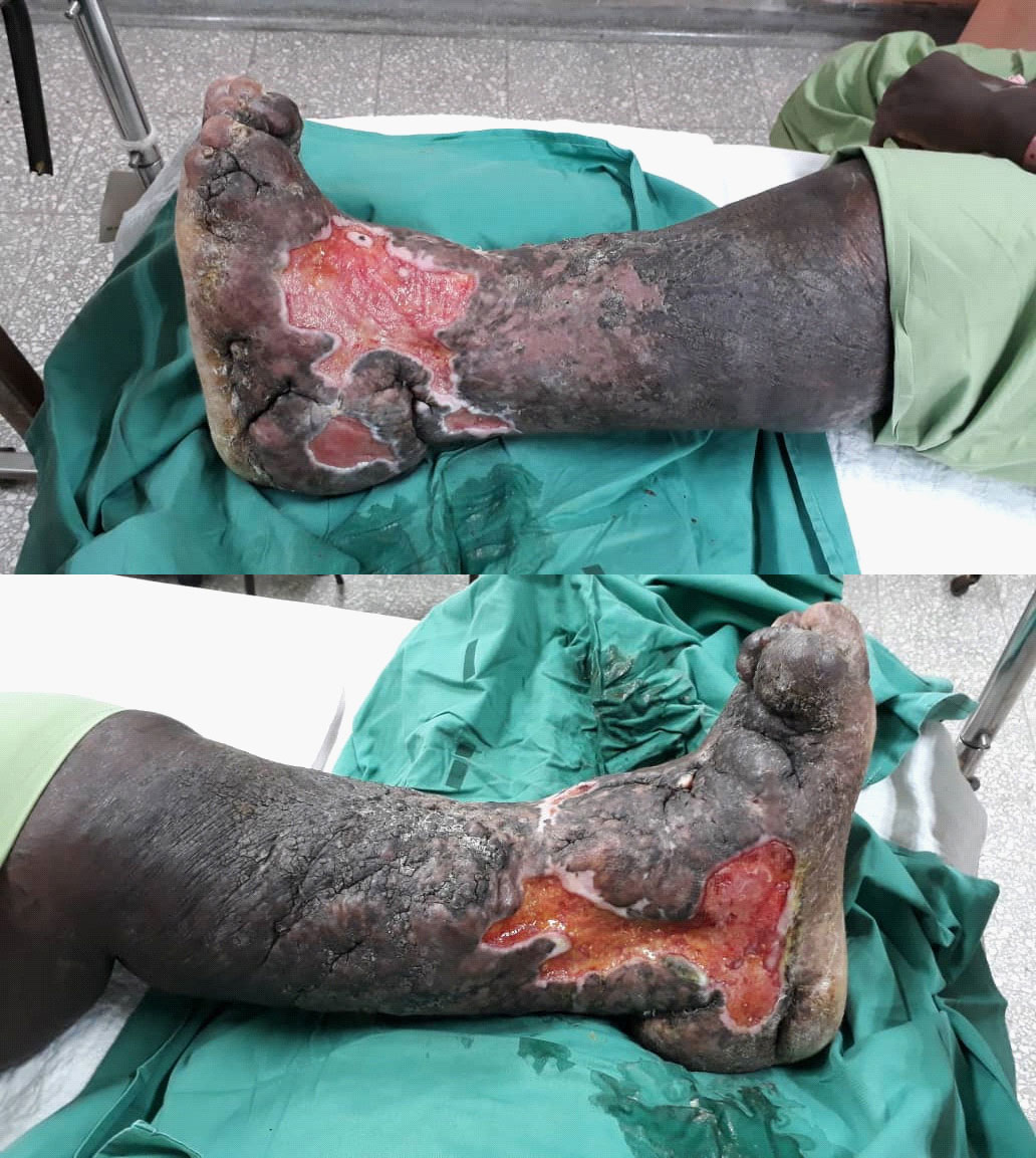
Upon admission, his blood pressure was 132/78 and his pulse rate of 82 bpm. Physical examination showed the left lower limb significantly swollen with lipodermatosclerosis and hyperpigmentation. His left calf is non-tender with a negative Homans sign. There is an ulcer at the gaiter's area measuring 12 X 15 cm, with sloping edges. The ulcer is involving almost the whole circumferential of the left ankle region. The wound is covered with pale granulation tissue and slough (Figure A & B). In addition, both the popliteal and dorsalis pedis arteries were not palpable due to thickened skin and edema. However, there is triphasic signal present over the bilateral popliteal, dorsalis pedis, and posterior tibial artery. Other systemic examinations are unremarkable.
Basic workup, including complete blood count, renal profile, chest X-ray, and coagulation profile, was within normal limits. Left lower limb duplex was done, showed no deep vein thrombosis, however, there are presences of incompetency of the left saphenofemoral junction, and patent left saphenopopliteal junction.
CT venogram was done showed significant compression of the left external iliac vein against the lumbar vertebrae by the left external iliac artery at the level of L5 vertebra. The vessel is, however, patent with no filling defect to suggest thrombosis. These findings on CT venogram are suggestive of May-Thurner syndrome.
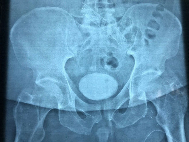
The patient underwent a left venogram and left external iliac vein stenting. Intraoperative findings showed narrowing of the left external iliac vein, which confirm the diagnosis of MTS. A stent measuring 16 mm X 100 mm was deployed in iliocaval junction with a good flow through the stent into IVC (Figure C). Post-surgery, he was started on heparin infusion and was overlapped with oral warfarin. For his venous ulcer, he was put on a vacuum dressing for 2 cycles and then was changed to four-layer compression dressing.
Discussion
May-Thurner syndrome is a rare cause of venous disease in the left lower limb. It is caused by compression of the left common iliac vein by the right common iliac artery. The artery appears to induce obstruction of the vein by its anatomical orientation with the entrapment of the left common iliac vein and by the extensive intimal hypertrophy of the vein from the chronic pulsation of the right common iliac artery. May-Thurner syndrome is estimated to occur in 2-5% of patients who undergo evaluation for lower extremity venous disorders [3].
A detailed history, physical examination, and diagnostic workup, including thrombophilia workup, should be performed to identify the risk factors for DVT. Patients with a known diagnosis of MTS should always have an evaluation for thrombophilia. Kolbel et al. [5] found that 67% of patients with chronic iliac vein occlusion or MTS have some form of thrombophilia. Our patient did not have any evidence of thrombophilia.
The patient may present with progressive disease, long standing, and with disabling complications [4]. It can cause leg edema, skin pigmentation, chronic varicosities, deep vein thrombosis, chronic venous stasis ulcers, or more serious complications, such as pulmonary embolism or phlegmasia cerulea dolens [3].
Color Doppler ultrasonography is the first-line method to diagnosed patients with common iliac vein disorder and to detect DVT. But this technique cannot accurately identify venous spurs or compression of the vein. The diagnostic test of choice is iliac venogram via femoral artery, to demonstrate the compression. Computed tomography venography (CTV) or magnetic resonance angiography (MRV) will show the offending overlying artery and show the presence of any extrinsic compression.
May-Thurner syndrome patients are commonly asymptomatic, and it is therefore unrecognized until symptoms develop. Treatment of symptomatic May-Thurner syndrome has evolved over the years from traditional open repair to less invasive endovascular repair. Treatment is aimed at clearing the thrombus present to prevent post-thrombotic syndrome and to correct the underlying compression of the left iliac vein. If left untreated, patients may develop debilitating post-thrombotic syndrome [7].
MTS is treated only when it is symptomatic. Previously open surgical procedures were done for the repair of MTS, but with the advancement of technology, less invasive endovascular repair has gained popularity. The principle of treatment is the removal of the clot with pharmacomechanical thrombolysis and mechanical thrombectomy to prevent post-thrombotic syndrome and to repair the anatomical defect with the use of stents and balloon venoplasty. Following placement of stents patients are treated with anticoagulants for at least six months to prevent in-stent restenosis. Kwak et al. showed that MTS-related DVT patients who had a metallic stent placed following thrombectomy had a primary and secondary patency rate of 95% and 100% at a 2-year follow-up [8]. A 73% recurrence rate was noted in patients with acute left-sided ilio-femora deep vein thrombosis when the underlying obstruction was not treated via stent placement [9].
Conclusion
A diagnosis of MTS should always be put as a differential diagnosis when presented with a case of unilateral DVT, especially in a younger age population. If the diagnosis is missed, it can have debilitating sequelae resulting from a post-thrombotic syndrome that will lead to significant morbidity and mortality. With early recognition and aggressive management, and availability of endovascular treatment, May-Thurner syndrome can be a well-managed disease.
Declarations
Ethics approval and consent to participate: This work adheres to the guidelines and principles of the Declaration of Helsinki and is in accordance with the Malaysian Good Clinical Practice (MGCP) 4th edition 2018. This research is also registered with the National Medical Research Register (NMRR), and ethics approval was obtained from the Medical Research and Ethics Committee (MREC) prior to the initiation of the study.
Consent for publication: Verbal consent was obtained from the patient.
Availability of data and materials: Not applicable.
Competing interests: The authors declare that they have no competing interests.
Funding: This study was conducted without any funding from any parties.
Authors' contributions: CJ collected the patient's clinic information, searched relevant works of literature, and wrote the manuscript. MAAM was the attending doctor of the patient. IZ & ZAA carried out critical revision and correction of the manuscript. All authors read and approved the final manuscript for submission and publication.
Acknowledgements: We would like to thank the Director-General of Health Malaysia for his permission to publish this article. We also would like to express our gratitude to the Surgical Department of Hospital Sultanah Aminah Johor Bahru, and those who had extended their help in contributing to this manuscript.
References
- R. May and J Thurner. The cause of the predominately sinistral occurrence of thrombosis of the pelvic veins. Angiology. 1957; 8: 419–427.
- May-Thurner syndrome: management by endovascular surgical techniques. Heniford BT, Senler SO, Olsofka JM, Carrillo EH, Bergamini TM Ann Vasc Surg. 1998; 12: 482-6.
- liocaval compression syndrome. Taheri SA, Williams J, Powell S, Cullen J, Peer R Nowakowski P, Boman L, Pisano S Am J Surg. 1987; 154: 169-72.
- Cil BE, Akpinar E, Karcaaltincaba M, Akinci D. Case 76: May-Thurner syndrome. Radiology. 2004; 233: 361–5.
- T. Kolbel, M. Lindh, M. Akesson, J. Wasselius, A. Gottsater, and K. Ivancev, "Chronic iliac vein occlusion: midterm results of endovascular recanalization," Journal of Endovascular Therapy. 2009; 16: 483-491.
- Brazeau NF, Harvey HB, Pinto EG, et al. May-Thurner syndrome: diagnosis and management. Vasa. 2013; 42: 96–105.
- Raffini L, Raybagkar D, Cahill AM, et al. May-Thurner syndrome (iliac vein compression) and thrombosis in adolescents. Pediatr Blood Cancer. 2006; 47: 834–838.
- H. S. Kwak, Y. M. Han, Y. S. Lee, G. Y. Jin, and G. H. Chung, "Stents in common iliac vein obstruction with acute ipsilateral deep venous thrombosis: early and late results," Journal of Vascular and Interventional Radiology. 2005; 16: 815–822.
- Mickley V, Schwagierek R, Rilinger N, et al. Left iliac venous thrombosis caused by venous spur: treatment with thrombectomy and stent implantation. J Vasc Surg. 1998; 28: 492–497.